Abdominal Anatomy Female - Stock Female Pelvis Normal Anatomy Illustrated Verdict / Media in category female human anatomy the following 144 files are in this category, out of 144 total.
byAdmin-
0
Abdominal Anatomy Female - Stock Female Pelvis Normal Anatomy Illustrated Verdict / Media in category female human anatomy the following 144 files are in this category, out of 144 total.. There is a common set of layers covering and forming all the walls: Its muscular part contributes to the lateral part of the abdominal wall. Anatomy and function of the female genitourinary system see online here get prepared for your anatomy exams: Jul 30, 2020 · the abdominal muscles also play a major role in the posture and stability to the body and compress the organs of the abdominal cavity during various activities such as breathing and defecation. The major muscles of the abdomen include the rectus.
The major muscles of the abdomen include the rectus. Skin, superficial fascia, muscles and associated fascia, and parietal peritoneum. In anatomy, the abdominal wall represents the boundaries of the abdominal cavity.the abdominal wall is split into the anterolateral and posterior walls. Anatomy of the female pelvis. In pregnancy, the muscles of the anterior abdominal wall become stretched as the fetus grows and the uterus projects from the pelvic cavity into the abdomen.
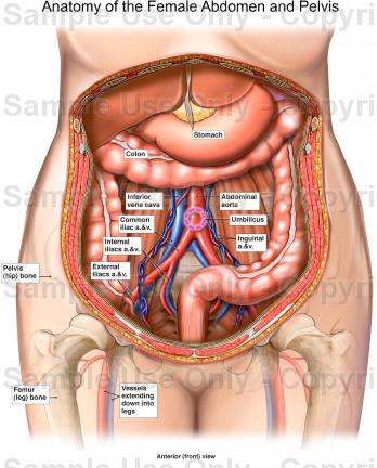
Human Female Anatomy Diagram Human Female Anatomy Diagram Human Anatomy Abdominal Anatomy Abdomen Mus Human Body Diagram Human Body Organs Human Body Anatomy from i.pinimg.com Here, you will find the most important information on the structure and function of the woman's internal and external sexual organs. Its muscular part contributes to the lateral part of the abdominal wall. In anatomy, the abdominal wall represents the boundaries of the abdominal cavity.the abdominal wall is split into the anterolateral and posterior walls. The upper boundary of the cervix is the level of the internal os, a narrowing of the uterus that is also referred to as the isthmus. It lies beneath the thoracic and abdominal skin, covering the internal abdominal oblique and anterior halves of the ribs and intercostal muscles. The muscles of the lower back, including the erector spinae and quadratus lumborum muscles, contract to extend and laterally bend the vertebral column. Anatomy of the female pelvis. There is a common set of layers covering and forming all the walls:
The muscles of the lower back, including the erector spinae and quadratus lumborum muscles, contract to extend and laterally bend the vertebral column.
It lies beneath the thoracic and abdominal skin, covering the internal abdominal oblique and anterior halves of the ribs and intercostal muscles. Here, you will find the most important information on the structure and function of the woman's internal and external sexual organs. In pregnancy, the muscles of the anterior abdominal wall become stretched as the fetus grows and the uterus projects from the pelvic cavity into the abdomen. There is a common set of layers covering and forming all the walls: The lower third of the uterus comprises the cervix. The muscles of the lower back, including the erector spinae and quadratus lumborum muscles, contract to extend and laterally bend the vertebral column. Skin, superficial fascia, muscles and associated fascia, and parietal peritoneum. Jun 17, 2021 · the abdominal wall is usually reinforced by a mesh to prevent further herniation before the incision is closed. Jul 30, 2020 · the abdominal muscles also play a major role in the posture and stability to the body and compress the organs of the abdominal cavity during various activities such as breathing and defecation. Pregnant women are susceptible to divarication of the recti. Jan 02, 2020 · abdominal obesity in females (15 f) b. These muscles help the body bend at the waist. Jan 24, 2018 · the muscles of the abdomen protect vital organs underneath and provide structure for the spine.
Here, you will find the most important information on the structure and function of the woman's internal and external sexual organs. Anatomy of the female pelvis. The anterolateral abdominal wall consists of four main layers (external to internal): May 31, 2021 · external abdominal oblique is the largest and the most superficial of the lateral abdominal muscles. Skin, superficial fascia, muscles and associated fascia, and parietal peritoneum.
Anatomy Of The Female Abdomen And Pelvis Cut Away View from healthiack.com There is a common set of layers covering and forming all the walls: Anatomy of the female pelvis. The anterolateral abdominal wall consists of four main layers (external to internal): Media in category female human anatomy the following 144 files are in this category, out of 144 total. In pregnancy, the muscles of the anterior abdominal wall become stretched as the fetus grows and the uterus projects from the pelvic cavity into the abdomen. Pregnant women are susceptible to divarication of the recti. Jan 24, 2018 · the muscles of the abdomen protect vital organs underneath and provide structure for the spine. Jun 17, 2021 · the abdominal wall is usually reinforced by a mesh to prevent further herniation before the incision is closed.
The major muscles of the abdomen include the rectus.
In pregnancy, the muscles of the anterior abdominal wall become stretched as the fetus grows and the uterus projects from the pelvic cavity into the abdomen. The muscles of the lower back, including the erector spinae and quadratus lumborum muscles, contract to extend and laterally bend the vertebral column. Pregnant women are susceptible to divarication of the recti. Its muscular part contributes to the lateral part of the abdominal wall. Jun 17, 2021 · the abdominal wall is usually reinforced by a mesh to prevent further herniation before the incision is closed. The major muscles of the abdomen include the rectus. Skin, superficial fascia, muscles and associated fascia, and parietal peritoneum. Anatomy of the female pelvis. The anterolateral abdominal wall consists of four main layers (external to internal): The lower third of the uterus comprises the cervix. In anatomy, the abdominal wall represents the boundaries of the abdominal cavity.the abdominal wall is split into the anterolateral and posterior walls. It lies beneath the thoracic and abdominal skin, covering the internal abdominal oblique and anterior halves of the ribs and intercostal muscles. There is a common set of layers covering and forming all the walls:
In anatomy, the abdominal wall represents the boundaries of the abdominal cavity.the abdominal wall is split into the anterolateral and posterior walls. Skin, superficial fascia, muscles and associated fascia, and parietal peritoneum. Here, you will find the most important information on the structure and function of the woman's internal and external sexual organs. The lower third of the uterus comprises the cervix. Jul 30, 2020 · the abdominal muscles also play a major role in the posture and stability to the body and compress the organs of the abdominal cavity during various activities such as breathing and defecation.
Female Anatomy Abdominal Adhesions Trialexhibits Inc from cdn.trialexhibitsinc.com Jan 24, 2018 · the muscles of the abdomen protect vital organs underneath and provide structure for the spine. Its muscular part contributes to the lateral part of the abdominal wall. Skin, superficial fascia, muscles and associated fascia, and parietal peritoneum. The major muscles of the abdomen include the rectus. In anatomy, the abdominal wall represents the boundaries of the abdominal cavity.the abdominal wall is split into the anterolateral and posterior walls. Pregnant women are susceptible to divarication of the recti. The lower third of the uterus comprises the cervix. Anatomy of the female pelvis.
Skin, superficial fascia, muscles and associated fascia, and parietal peritoneum.
May 31, 2021 · external abdominal oblique is the largest and the most superficial of the lateral abdominal muscles. There is a common set of layers covering and forming all the walls: Jun 17, 2021 · the abdominal wall is usually reinforced by a mesh to prevent further herniation before the incision is closed. The muscles of the lower back, including the erector spinae and quadratus lumborum muscles, contract to extend and laterally bend the vertebral column. In anatomy, the abdominal wall represents the boundaries of the abdominal cavity.the abdominal wall is split into the anterolateral and posterior walls. The lower third of the uterus comprises the cervix. Pregnant women are susceptible to divarication of the recti. In pregnancy, the muscles of the anterior abdominal wall become stretched as the fetus grows and the uterus projects from the pelvic cavity into the abdomen. Skin, superficial fascia, muscles and associated fascia, and parietal peritoneum. It lies beneath the thoracic and abdominal skin, covering the internal abdominal oblique and anterior halves of the ribs and intercostal muscles. Media in category female human anatomy the following 144 files are in this category, out of 144 total. The anterolateral abdominal wall consists of four main layers (external to internal): These muscles help the body bend at the waist.
It lies beneath the thoracic and abdominal skin, covering the internal abdominal oblique and anterior halves of the ribs and intercostal muscles abdominal anatomy. Jan 02, 2020 · abdominal obesity in females (15 f) b.